Researchers create ‘Mini-Brains’ in lab to study neurological diseases
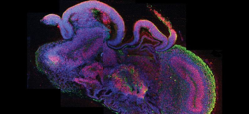
Researchers at the Johns Hopkins Bloomberg School of Public Health, with the participation of a Greek researcher, announced at a conference for the American Association for the Advancement of Science, they have successfully developed “mini-brains” made up of neurons and cells of the human brain that are capable of replicating some of its functionality. The researchers hope that their tiny balls of brain cells will change the way new drugs are tested for effectiveness and safety, obviating the need for animal trials.
Georgia Makri is the Greek researcher and Postdoctoral fellow in the Neurology Department of John Hopkins.
“Ninety-five percent of drugs that look promising when tested in animal models fail once they are tested in humans at great expense of time and money,” study leader Thomas Hartung, Professor and Chair for Evidence-based Toxicology at the Bloomberg School, said in a press release. “While rodent models have been useful, we are not 150-pound rats. And even though we are not balls of cells either, you can often get much better information from these balls of cells than from rodents.”
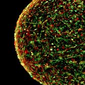
These neuronal clusters are only about 350 micrometers in diameter, approximately the size of the eye of a housefly. To grow these brains, Hartung and his colleagues made use of induced pluripotent stem cells, adult cells that have been genetically reprogrammed to an embryonic stem cell-state.
Hartung and his colleagues used skin cells from healthy adults as their starting point for the mini brains. These cells were stimulated to grow into brain cells and were then cultivated for eight weeks. After eight weeks, the mini-brains developed four types of neurons and two types of support cells, known as astrocytes and oligodendrocytes. The latter support cell creates myelin, a fatty white substance which insulates the neuron’s axons and allows them to communicate faster.
The researchers were able to watch the myelin growing and were able to record spontaneous electrophysiological activity in the mini-brains. In order to test them out, the team placed the brains on an electrode array and listened to the neuron’s’ spontaneous electrical activity as various test drugs were added.
“We don’t have the first brain model nor are we claiming to have the best one,” said Hartung. “But this is the most standardized one. And when testing drugs, it is imperative that the cells being studied are as similar as possible to ensure the most comparable and accurate results.”
According to Hartung, hundreds of mini-brains can be grown from the same batch of cells, with up to one hundred in a single petri dish. In addition to being used to study pharmaceuticals, they can also be used to study neurodegenerative diseases such as Parkinson’s and Alzheimer’s, as well as viral infections, trauma and, stroke. Hartung is currently in the process of applying for a patent for his mini-brains and plans to begin producing them commercially in 2016 through an entity called ORGANOME.


