Aims to explore the predictive biomarker potential of circulating tumor cells
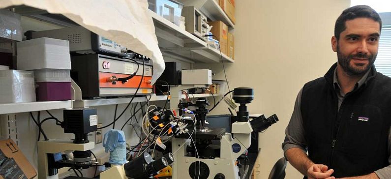
Dimitrios Papageorgiou is a Postdoctoral Associate at MIT’s (Massachusetts Institute of Technology) Department of Materials Science and Engineering.
Dimitris specialized on the interfacial phenomena of the interaction of electric fields and liquids on functional surfaces, as well as the integration of those in novel lab-on-a-chip diagnostic devices.
Supported by the TFP, Dimitris has pushed the technological development of a microfluidic platform using surface acoustic waves that can effectively filter out circulating tumor cells (CTCs) from peripheral blood samples of cancer patients. He envisions that acoustophoretic CTC isolation will play a key role in ongoing and future studies that aim to explore the predictive biomarker potential of CTCs, and personalized therapy decision making including novel immunotherapeutic approaches.
He was born on January 19th 1983 in Athens, Greece. He studied Physics at the National and Kapodistrian University of Athens (2000-2006), where he also obtained his Master’s Degree in Microelectronics (2007-2009). In 2013 he earned his PhD from the School of Chemical Engineering of the National Technical University of Athens. From 2007 to 2010 he worked as a physicist at the National Center for Scientific Research (NCSR) “Demokritos” in Athens, Greece.
Papageorgiou participated in a new study, in which scientists have developed a new system to classify the shapes of red blood cells in a patient’s blood using a computational approach known as deep learning. The findings, published in PLOS Computational Biology, could potentially help doctors monitor people with sickle cell disease. The scientists were led by another Greek researcher, George Karniadakis.
To automate the process of identifying red blood cell shape, they developed a computational framework that employs a machine-learning tool known as a deep convolutional neural network (CNN). The new framework uses three steps to classify the shapes of red blood cells in microscopic images of blood. The researchers validated their new tool using 7,000 microscopy images from eight sickle cell disease patients.
The research team plans to further improve their deep CNN tool and test it in other blood diseases that alter the shape and size of red blood cells, such as diabetes and HIV. They also plan to explore its usefulness in characterizing cancer cells.


