Greek researchers created support system for the early diagnosis of malignant melanoma
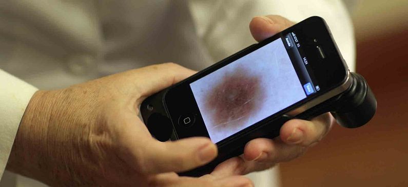
A team of Greek researchers from the Technological Educational Institution (TEI) of Athens developed a self-examination platform/application able to adjust in commercial smartphone or handheld devices for facilitating early stage detection of melanoma.
Melanoma is considered the most serious and most commonly fatal form of skin cancer. Over the past 4 decades, the incidence of melanoma increased by 800% among young women and 400% among young men. Early stage detection of melanoma (one of the most common cancers today) is of major significance for increasing chances of long term survival of affected patients.
MARK1 project is a co-funded GREECE-ISRAEL research cooperation that aims in developing a self-examination platform/application able to adjust in commercial smartphone or handheld devices for facilitating early stage detection of melanoma. MARK1 will incorporate image processing, image analysis and pattern recognition techniques for mole identification, analysis and classification. The application will be linked with the medical specialist who will be able to advice regarding the urgency for a medical examination. In this way, MARK1 aims in early and accurate screening of melanoma by an easy to use technology accessible by general public, in order to enable fast, offline supervision of suspected moles by the health care specialist for detecting the disease at its early stages, when it is more vulnerable to available treatments.
The team’s main objective is to present the conceptual architecture of a platform that can address the need for early and accurate detection of skin lesion through a screening solution that will be easily accessible to the general public with the guidance, supervision and inspection of the primary care physician.
The system was designed on the GPU card (GeForce 580GTX) using CUDA programming framework and C++ programming language. Results showed that the designed system discriminated benign from malignant moles with 88.6 % accuracy employing morphological and textural features. The proposed system could be used for analysing moles depicted on smart phone images after appropriate training with smartphone images cases. This could assist towards early detection of melanoma cases, if suspicious moles were to be captured on smartphone by patients and be transferred to the physician together with an assessment of the mole’s nature.
The Medical Image and Signal Processing (MEDISP) laboratory is part of the Department of Biomedical Engineering of the Technological Educational Institution (TEI) of Athens Greece. MEDISP’s main objective is to research and develop new methods and applications for Medical Image Processing, Image Analysis and Pattern Recognition, Radiation Detectors (Image Formation), Medical Statistics and Medical Informatics to assist in the solution of clinical problems.


