The Greek who makes cancer tissues glow
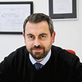
The path towards the more effective removal of tumors has been paved by Vasilis Ntziachristos, head of the Institute of Biological and Medical Imaging at the Technical University of Munich and the Helmholtz Center in Germany.
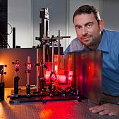
Prof. Ntziachristos´s area of research encompasses the development of new methods and devices for biological and medical imaging focusing on innovative non-invasive approaches that visualize previously unseen physiological and molecular processes in tissues. His research also aims to translate these methods to advance biological discovery, accelerate drug development and offer new efficient methods for diagnostics and theranostics.
Prof. Ntziachristos studied electrical engineering at Aristotle University in Thessaloniki, Greece and received his Master’s and Doctorate degrees from the Bioengineering Department of the University of Pennsylvania. He served as assistant professor and director of the Laboratory for Bio-Optics and Molecular Imaging at Har-vard University and Massachusetts General Hospital. His current professorial chair is closely linked with the Institute of Biological and Medical Imaging at the Helmholtz Zentrum München, of which Prof. Ntziachristos is director.
In 2012, Ntziachristos’ team, consisting of Georgios Themelis and Athanasios Sarantopoulos, published its report in the “Nature Medicine” journal. The researchers injected a substance resembling folic acid into patients , thereby making cancer tissues glow under the light of a special camera. This allows surgeons to separate tumors from healthy cells during operations. According to existing data, scientists using this method can spot an average of 34 tumors in each patient, compared to an average of seven tumors that could be identified using previous methods. Vasilis Ntziachristos refers to this discovery as a “true leap of progress in surgical imaging”, noting that “until now, we could only rely on the human eye to find carcinogenic tissue, or non-specific dyes that would color the vascular tissue as well as particular cancer cells. Now we are going after precise molecular signals and not simply physiology. This provides more accuracy and more certainty for clinicians to remove cancerous cells in real time during surgery».
Read also:


