The biophysicist who tries to make cancer detection a lot easier
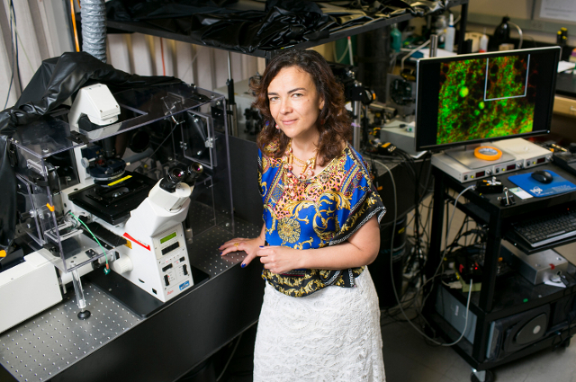

Irene Georgakoudi is an Associate Professor of Biomedical Engineering at Tufts University. She has been working on the use of lasers for therapeutic and diagnostic applications since her undergraduate years. She is developing a new optical technique that may, one day, make screening for cervical cancer faster, easier, and more precise. The tool is a high-resolution microscope that makes it possible to visualize and analyze individual cells instantaneously using very fast pulses of light.
Irene Georgakoudi got her B.A. in Physics with high honors at Darmouth College, Hanover (1989-1993) and in 1998 she got her Ph.D. in Biophysics at the University of Rochester (School of Medicine and Dentistry, NY) where she specialized in Photodynamic Therapy of Cancer. Through her academic career she has work in several famous insitutions as a Research Scientist (Massachusetts Institute of Technology, Harvard Medical School, Tufts University and École Polytechnique, Laboratoire d’Optique et Biosciences, France).
Every year, millions of women undergo a Pap smear to look for early cervical cancers or pre-cancerous changes in the cervix. This test was introduced in the late 1940s and has helped to significantly reduce mortality from cervical cancer in the decades since. This test, however, has limitations. For example, a specialist must use his or her judgment to visually determine if cells look abnormal, which can lead to false positives. Because of this, women often need a biopsy.
“This can cause a lot of unneeded cost and stress”, says Irene Georgakoudi, who is working with Karl Munger, Ph.D., a Tufts University virologist, on a more reliable and less invasive way of detecting cancers, such as cervical, early. Georgakoudi’s new tool, which she is developing with the help of funding from the American Cancer Society, would allow for an automated analysis of results, removing “human subjectivity from the interpretation process.
“The goal for the researchers in my field is to develop techniques that basically allow us to identify microscopic changes at an early stage that are both more sensitive and more specific than current tools allow, so doctors won’t need to biopsy tissue if there is a good chance the tissue is not going to be abnormal.”
Georgakoudi’s tool is a high-resolution microscope whose magnification power far outstrips that of a colposcope, the tool currently used to examine the cervix to look for cancer. “Instead of visualizing the surface of the cervix at 6 to 15 times magnification we are looking at magnifications that are on the order of 25 to 60 times, so we can really visualize individual cells”.
Georgakoudi’s team used an noninvasive microscopy technique that exploits NADH fluorescence enables researchers to observe how mitochondria alter their shape and arrangement in human tissue.Patients could one day get their skin examined under a special microscope and, in just a few minutes, know whether they have cancer, according to this study published on November 30,2016.
She has received many awards and honors for her distinguished work (in fact she has been inducted in The American Institute for Medical and Biological Engineering (AIMBE) for outstanding contributions to the development of optical methods for cancer diagnosis and tissue engineering applications). She has co-authored nearly 50 peer-reviewed articles and book chapters and has presented her work at national and international meetings. She has served on the Board of Directors of the Optical Society of America.